Industrial CT – A Powerful Tool for Archaeologists, Art Experts, Museums, and More
Industrial computed tomography (CT) is a highly advanced and non-destructive technique that allows for the examination of the internal structure of objects in three dimensions. The rapid advancements in CT technology have led to a surge in the use of industrial CT scanners across various sectors, expanding both their applications and the industries they serve.
An emerging and intriguing trend is the increased use of CT scanning in fields beyond the traditional industrial applications. We have received a growing number of CT scanning requests from archaeologists, art experts, and museums. In this article, we highlight one particularly fascinating application: the CT scanning of a Merovingian belt buckle, which was imaged immediately after being excavated, still encased in clay.
Acquisition of the Merovingian belt buckle
A key advantage of CT technology is its non-destructive nature. It does not heat the specimen and is effective with any surface, shape, color, or material (within certain density or thickness limits that X-rays can penetrate). In this case, the belt buckle was scanned in its original state, still encased in the clay from the excavation site.

In operation, CT generates X-ray photons that either travel through the object or are attenuated by it. The photons that penetrate the object enter the imaging detector, carrying with them detailed information (see image below). The degree of photon attenuation depends on the amount of energy applied and the thickness or density of the material that the photon pass through.
On this specific sample, 1800 images have been acquired (1 image every 0.2 degree). Each image is 8 Megapixels and is also averaged and filtered to reduce noise. The 2D digital images taken during this step are saved directly into a single folder which will be used in the next step of the CT process.

Reconstruction and Visualization
Once the acquisition process of the CT scan is completed, CT calibration and CT reconstruction algorithms are used to reconstruct the 3D CT volume. These 3D images are made of Voxels (three dimensional Pixels), and with the use of visualization software the 3D volume can be manipulated in real time. Now it is possible to slice through anywhere inside the object, inspect and look for key features and take accurate measurements.
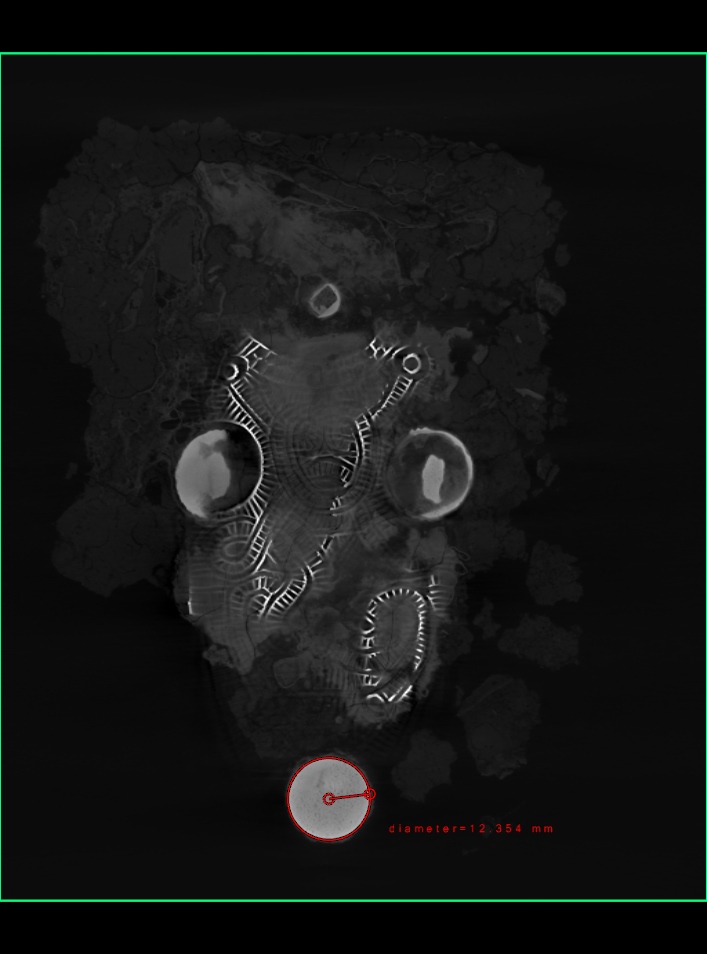
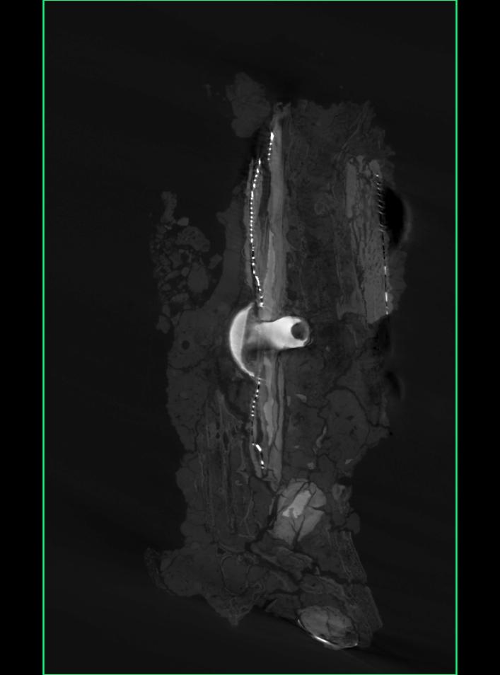
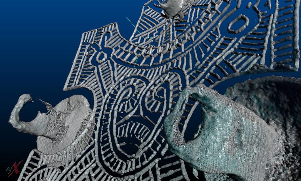
The 3D CT reconstruction, typically consisting of millions of voxels, can be converted into a surface model. The operator can set a threshold value of radiodensity using edge detection algorithms, allowing a 3D model (polygon mesh) to be generated and displayed on screen. Different models can be created by adjusting radiodensity thresholds, with each component of an assembly represented by a distinct color.
Multiple models can be constructed from various different radiodensity thresholds, therefore allowing different colors to represent each component of an assembly.
In this specific application, the clay and the metallic belt buckle are represented in different grey colors. After segmentation and surface reconstructions, those 2 components can be displayed in 2 different colors.
Additionally, the histogram can be segmented to isolate a single component, such as the higher-density metallic buckle.
The resolution of the 3D surface model depends on the number of voxels generated during CT reconstruction, which in turn is influenced by the size of the object being scanned. Industrial micro-CT scanners can achieve resolutions hundreds of times greater than those used in medical imaging, with 3D models often comprising thousands to up to 50 million polygons. The output formats (such as point clouds or STL files) are compatible with rapid prototyping machines, allowing for direct use in 3D printing applications.
Advantages and Applications
Over the years, industrial CT technology has evolved rapidly, becoming a competitive option for 3D scanning. Its ability to provide highly accurate internal details of an object, without destruction, and to assess the state of preservation before any costly or time-consuming restoration work, is unparalleled.
Industrial CT scanning offers numerous advantages: there are no shaded areas, it works with virtually any shape or surface, and almost any material (as long as its density or thickness permits adequate X-ray penetration). Minimal or no post-processing is required, and the resolution of both internal and external features is exceptional.
Overall, 3D CT technology is unmatched in its capabilities, making it an invaluable tool for a wide range of industries, research labs, archaeologists, art experts, museums, and more. It opens up new possibilities for understanding and preserving cultural heritage, as well as optimizing industrial processes and product development.
Talk to Our Experts