Computed Tomography for Firearms and Munitions
Computed Tomography for Military and Defense
In today’s competitive global market, engineers are faced with the ever growing challenge of balancing the functionality, complexity, reliability, safety, aesthetics and cost of a product’s design while bringing it to market faster than anyone has done before. Digital radiography (DR) and Computed Tomography (CT) give the modern engineer tools like no other. Analysis, reverse engineering, model building and communication all benefit from DR &CT X-ray technology.
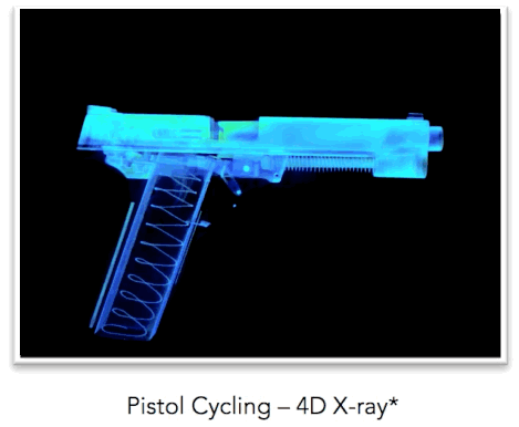
Computed Tomography is the capture of X-ray photons with electronic circuit panels. Directly converting X-ray photon energy into pixel data brings the economy of speed and the flexibility of digital database analysis to the engineers. Gone is the need for harsh chemicals, development labs and environmentally controlled archive rooms. This digital database approach to X-ray image processing has opened the doors to motion X- rays, high resolution 3D X-rays and even motion 3D X-rays, now called 4D X-rays. Assessing the quality of a system’s internal components can be difficult after assembly. In some cases an engineer may have no option but to destructively sample a small number of systems to make quality predictions about a larger population. The cost of finished goods lost in destructive quality auditing is typically absorbed by the manufacturer in the form of overhead and is passed along to the consumer in the final price. With the application of computed tomography, an engineer may have the ability to non-destructively assess a system’s integrity in ways never before available.
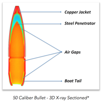
As an excellent example, let’s consider the challenge of assessing a bullet’s integrity without dismantling it. A bullet is a precision part and may be composed of multiple components. Traditionally, measuring jacket uniformity, core conformity and void proclivity meant cutting the bullet in half and looking at it under a microscope. Sectioning the bullet in this manner is labor and time intensive, as well as being potentially destructive of the finished product. The biggest drawback to this type of destructive test is sectioning in the wrong location, causing a potential defect to be missed.
If a bullet’s component integrity and geometry is assessed via 3D CT X-ray, the scan and analysis may be performed in a matter of minutes. Jacket uniformity, core conformity and void proclivity can be assessed for the entire bullet, not just along a single section, greatly reducing the chance of overlooking a defect. Utilizing 3D X-ray analysis the bullet would still be a viable finished good. This opens the possibility for increased quality control sample sizes and increased production capability knowledge while reducing overhead costs.The precision of a 3D X-ray scan can also significantly aid in the study, reverse engineering and understanding of a competitor’s complex product. For example, telescopic rifle scopes contain many small, precision parts. Most rifles scopes are designed to allow the user to make very fine and precise alignment changes to the internal glass of the scope. This is done to adjust magnification, focus and target alignment with respect to the shooter’s eye and bullet trajectory. Once those fine adjustments have been made to the scope, the adjustments must hold up under the recoil forces of the rifle shot after shot. It is easy to see why precision rifle scopes are incredibly challenging to design and manufacture. This is why precision rifle scopes often cost more than the firearm to which they are attached.
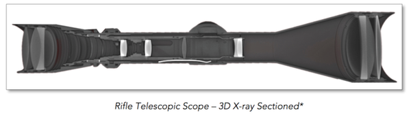
Imagine the time savings if an engineer who wanted to study a competitor’s scope could examine each part in its entirety while keeping its physical relationships to all the other parts intact? By implementing computed tomography, a scope could be accurately memorialized – part counts taken, measurements made, part-to-part relationships established and component motion captured. A digital database of a scope can be made in a matter of hours, rather than the days usually associated with dismantling a scope and measuring all of the individual parts while laying out their relationships.
Model building also benefits from computed tomography. If a lens of a scope being studied has a unique shape (non parabolic), accurately capturing the shape may be difficult. An engineer could employ a coordinate measurement machine (CMM) and create a cloud of data points by touching many locations on the lenses’ surfaces. The point cloud could then be imported into a computer aided drafting (CAD) program and the lenses’ surfaces could be interpolated and used to create a model. Conceivably, a more efficient method would be to create a 3D X-ray scan of the lens and export its surfaces directly to a model file. Once the model file has been created, most CAD software packages can examine and manipulate the model in almost anyway the operator desires.
One arena in which DR & CT technology are playing an increasingly important role is product liability litigation. If an accusation of a defective product has been made, it may be possible to determine if a defect exists without the associated arguments of alteration that are occasionally made after disassembly. In litigation, the preservation of evidence is paramount.
The precision and nondestructive nature of computed tomography makes it a strong fit for evidence evaluation. Objects are typically not heated, cooled or subjected to magnetic fields when scanned. Therefore, products and their components may be memorialized without damaging or altering them in many cases.
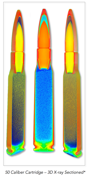
In the world of litigation, getting permission to perform an exam on a suspect product can be very complicated if the exam may alter the physical evidence in any way. Measuring the pressure produced by a firearm cartridge when it is discharged is an excellent example of such a test. When a cartridge is discharged, typically the primer and propellant/powder are consumed. The brass shell and the bullet may also significantly distort. Therefore, the discharge of ammunition to assess the produced maximum pressure would be considered destructive of that particular cartridge.
Assume you are an analyst and 1,000 rounds of suspect firearm ammunition needs to be pressure tested, and the court will only allow X rounds of that ammunition to be consumed in pressure testing. Which X rounds do you choose? Employing 3D X-ray scanning technology may allow the component integrity of all 1,000 rounds to be nondestructively assessed. Knowing the makeup of all 1,000 rounds could show they are all physically similar and suggest destructively testing a random sample of X units may provide an acceptable pressure characterization. Conversely, a 3D X-ray assessment of all 1,000 rounds could reveal physical anomalies in specific rounds and testing those rounds would insure potentially critical evidence is not missed due to random selection. In short, the judicial use of computed tomography could substantially build confidence in the foundation upon which an analysis is built.
Getting to the facts of a products design and manufacture is where computed tomography in its many forms shines. Whether it is in the fields of product development, testing, quality control, reverse engineering, model building or litigation, digital radiography is opening the doors traditionally closed to engineers. At the forefront of this industrial X-ray revolution is North Star Imaging, Inc. (4nsi.com). NSI manufactures the DR and CT systems, writes the software, trains the operators and provides consulting services. NSI’s synergy of products and services enables companies to see their finished goods in ways their designers may never have imagined.https://www.youtube.com/embed/QpRY1ErSxs0
As engineers, analysts and consultants, gathering accurate data and interpreting it are only two of the challenges we face. Communicating the data and its meaning in a manner easily understood is equally as challenging. Newspaper editor Arthur Brisbane was quoted in a 1911 article as saying, “Use a picture. It’s worth a thousand words”. For me, the use of computed tomography has simplified the initial course of action for many challenges to a simple statement of four words, “Let’s take a look.”
